
 |
Parkinson’s disease has become better known to the general public through awareness of the disease in famous people such as Michael J. Fox. Parkinson’s disease is a progressive neurodegenerative disorder of the central nervous system characterized by a loss of dopaminergic neurons in the substantia nigra in the midbrain. The hallmark symptoms are bradykinesia, rigidity, and resting tremor. However, it is commonly known that disturbances of sleep also may be seen in these patients. People with Parkinson’s disease suffer from insomnia, excessive daytime sleepiness, sleep attacks, nightmares, periodic limb movement in sleep, restless legs syndrome, obstructive sleep apnea syndrome, and REM sleep behavior disorder.1
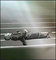
REM sleep behavior disorder is a very interesting disorder of REM sleep. In a nutshell, people are normally paralyzed during REM sleep, but patients with REM sleep behavior disorder act out their dreams. It may be that the actions witnessed during such an event may correlate relatively well with the patient’s later description of what was occurring in their dream. These movements can be somewhat abrupt and even violent and may result in injury to the patient or their bed partner.
IDENTIFYING REM SLEEP BEHAVIOR DISORDER PATIENTS
Which patients are likely to have REM sleep behavior disorder? A study of patients with the disorder seen at the Mayo Sleep Disorders Center demonstrated that 87% were male and that the mean age of onset was 60.9 years (range 36-84 years). Acting out dreams is interesting, but it may also be problematic. During episodes of acting out dreams, 32% of these patients had injured themselves and 64% had assaulted their spouses. In 87% of these cases, dream content frequently involved defense of the sleeper against attack. Neurological disorders were present in 57% of these patients, the most common being Parkinson’s disease. Acting out dreams in an otherwise symptom-free patient may sometimes be a harbinger that worse is yet to come as REM sleep behavior disorder actually developed before parkinsonian symptoms in 52% of the patients.2 In support of this, neuroimaging studies may show a reduction of striatal dopamine transporters even before the diagnosis of Parkinson’s disease becomes evident. A study of single photon emission computed tomography (SPECT) scans performed in normal controls and in patients with REM sleep behavior disorder sheds some light on this. Not surprisingly, the signal in the striatum was normal in the control subjects. There was a significant decrease in signal in patients with subclinical REM sleep behavior disorder, a further decrease in signal in patients with clinically manifest REM sleep behavior disorder, and an even further decrease in signal in patients with REM sleep behavior disorder who also had a diagnosis of Parkinson’s disease.3 Another study of patients with a history of REM sleep behavior disorder not yet associated with a neurological disorder demonstrated that 45% of these patients developed Parkinson’s disease after a mean of 11.5 years from the reported onset of REM sleep behavior disorder.4 This data flows nicely with the concept that the degree of nigrostriatal degeneration correlates with the degree of symptoms, and that the diagnosis of Parkinson’s disease can come either before or after the onset of REM sleep behavior disorder.
 |
The real paradox here is that normal subjects do not act out their dreams during REM sleep, while patients with REM sleep behavior disorder who also have Parkinson’s disease do not move normally while awake. However, an incredibly thought-provoking study of patients with both Parkinson’s disease and REM sleep behavior disorder demonstrated that the movements seen during an episode of acting out a dream may actually appear to be relatively normal, at least in the sense of not demonstrating the expected tremor, rigidity, and bradykinesia that defines the motor movements of Parkinson’s disease seen during wakefulness. Bed partners of these patients believed that while acting out a dream, the motor movements of the patients were improved in 87%, speech was better in 77%, and facial expression was normalized in 47% of the patients. The video-monitored purposeful movements during REM sleep were also surprisingly fast, ample, coordinated, and symmetrical, without obvious sign of parkinsonism. The movements were, however, sometimes jerky, violent, and repetitive. How is it that there may be a difference in the liberated movements of the dreaming patient with Parkinson’s disease compared to what may be attained by the same patient during normal wakefulness? The pyramidal motor system is the one we use during normal volitional motor movements, and the extrapyramidal pathway is another motor pathway that is less well understood. The mechanism behind the apparent restored motor control during REM sleep is unclear, but the authors proposed that parkinsonism may disappear during REM sleep through a disconnect between pyramidal and extrapyramidal motor systems.5
CONTROLLING THE MOTOR PATHWAY
Can the motor pathway used by a patient with Parkinson’s disease while acting out a dream somehow become called into action during normal wakefulness? It is at least conceivable that this observation may someday lead to the use of such pathways during wakefulness, thus perhaps restoring an improved motor state of decreased motor rigidity, tremor, and bradykinesia. The most obvious possible intervention that would at least have the potential of disabling certain motor pathways while enabling others would be deep brain stimulation, a common treatment modality used in patients with Parkinson’s disease today. The device really looks somewhat like a pacemaker that is placed in the subcutaneous fat below the collar bone just as a pacemaker is. The difference is that the wires are tunneled beneath the skin up the neck, travel through a small hole in the skull, and terminate deep in the brain where they stimulate deep nuclei. The most common nuclei stimulated by these devices are either the subthalamic nucleus or the internal globus pallidus, both deep within the brain.
DEEP BRAIN STIMULATION
How does deep brain stimulation work? The truth is that it is not completely understood, but it certainly helps these patients. These improvements are strongly correlated with improvements in motor function, primarily with regard to the freezing of normal motor movement known as bradykinesia.6 The primary improvement in patients with bilateral subthalamic nucleus stimulators may be most obvious during the “off” medication time, that is, when the symptoms of rigidity would normally return following the metabolism and decreased blood levels of the dopaminergic agonist medication just prior to the next dosing of the drug.7 Improvements in quality of life following bilateral deep brain stimulation of the subthalamic nucleus are maintained in the long term. In patients with advanced Parkinson’s disease who underwent bilateral deep brain stimulation of the subthalamic nucleus, sustained improvement in motor function has been demonstrated to persist for 2 years after the procedure.8 A 5-year prospective study of patients with bilateral stimulation of the subthalamic nucleus demonstrated improved motor function even while off medication by up to 54% and improved activities of daily living by 49%.9 However, following this time period, symptoms may again worsen as the natural disease progression worsens.10
[registe
r] [/register] [register]Want more information about REM sleep behavior disorder? Subscribe to Sleep Report to receive the latest research findings. [/register]
Not only do deep brain stimulators improve motor movements, but interesting improvements may also be seen during sleep. Patients with subthalamic nucleus deep brain stimulation may experience an increase in total sleep time, improved sleep efficiency, and an increase in slow wave sleep and REM sleep.11 The beneficial effects on both motor movement and sleep by deep brain stimulation of the subthalamic nucleus may persist through time. Three months after placement of a subthalamic nucleus deep brain stimulator, sleep study findings may include an increase in total sleep time, an increase in the longest period of uninterrupted sleep,12 an increase in slow wave sleep, and a reduction of periodic limb movements. Improvement in the subjective state of the patient during daytime wakefulness may be seen, likely due to a reduction of sleep fragmentation.13 For these patients, subthalamic nucleus deep brain stimulation may provide an improvement in quality of life, reduce requirements for medication, and possibly enhance mental flexibility.14 However, not all sleep abnormalities of Parkinson’s disease improve with deep brain stimulation. For those patients with obstructive sleep apnea, the apnea-hypopnea index may show no improvement. Also, the motor movements of REM sleep behavior disorder do not usually improve.15 That is, deep brain stimulation of the subthalamic nucleus does not restore the normally seen paralysis of REM sleep.
How much better do these patients feel once their stimulators are surgically implanted? High-frequency deep brain stimulation of the subthalamic nucleus improves the motor symptoms of Parkinson’s disease and may provide subjective feelings of well-being, euphoria, an increase in motivation, and a decrease in fatigue.16 Although the subthalamic nucleus is not the pleasure center, sometimes the results may be a little too good. There is one case report of a patient who had benefited from bilateral subthalamic nucleus deep brain stimulation for Parkinson’s disease but who also then developed acute and reproducible manic behavior when stimulation occurred in the substantia nigra.17
The subthalamic nucleus is the most often targeted deep brain nucleus in patients with Parkinson’s disease, but what does the subthalamic nucleus have to do with REM sleep? Deep brain stimulators are, of course, primarily used for stimulating deep nuclei of the brain, but they also have the ability to record from neurons deep in the brain that surround the tip of the electrode. Interestingly, neuronal firing recorded by a deep brain stimulator in the subthalamic nucleus may show neuronal firing that corresponds with the rapid eye movements of REM sleep. The neurons usually fire just slightly after the actual eye movements, and thus, although there is no evidence that the subthalamic nucleus is responsible for driving eye movements, it may have a role in sensory feedback of the eye movements of REM sleep.18
Would it really be possible for deep brain stimulation to induce an extrapyramidal motor pathway that could restore or at least improve mobility in a patient with Parkinson’s disease? Interestingly, there is a case of REM sleep behavior disorder that occurred suddenly following the placement of a left subthalamic deep brain stimulator. The authors believed that the stimulator caused a microlesion in or near the upper part of the pars compacta of the substantia nigra, which was likely responsible for the subsequent onset of REM sleep behavior disorder in this patient.19 Although this does not necessarily mean that a similarly placed stimulator could bring about more normal motor movements during wakefulness, it may be a clue headed in the right direction.
Timothy J. Walter, MD, DABSM, and Uma Marar, MD, DABSM, are sleep medicine physicians operating the American Academy of Sleep Medicine-accredited practice Capitol Sleep Medicine in Grove City, Ohio. They can be contacted at [email protected].
REFERENCES
- Boczarska-Jedynak M, Opala G. Sleep disturbances in Parkinson’s disease. Neurol Neurochir Pol. 2005;39:380-388.
- Olson EJ, Boeve BF, Silber MH. Rapid eye movement sleep behavior disorder: demographic, clinical and laboratory findings in 93 cases. Brain. 2000;123:331-339.
- Eisensehr I, Linke R, Tatsch K. Increased muscle activity during rapid eye movement sleep correlates with decrease of striatal presynaptic dopamine transporters. Sleep. 2003;26:507-512.
- Iranzo A, Molinuevo JL, Santamaria J, et al. Rapid-eye-movement sleep behaviour disorder as an early marker for a neurodegenerative disorder. Lancet Neurol. 2006;5:572-577.
- De Cock VC, Vidailhet M, Leu S, et al. Restoration of normal motor control in Parkinson’s disease during REM sleep. Brain. 2007;130(Pt 2):450-456.
- Lyons KE, Pahwa R. Long-term benefits in quality of life provided by bilateral subthalamic stimulation in patients with Parkinson disease. J Neurosurg. 2005;103:252-255.
- Liang GS, Chou KL, Baltuch GH, et al. Long-term outcomes of bilateral subthalamic nucleus stimulation in patients with advanced Parkinson’s disease. Stereotact Funct Neurosurg. 2006;84(5-6):221-227. Epub 2006 Oct 23.
- Kleiner-Fisman G, Fisman DN, Sime E, Saint-Cyr JA, Lozano AM, Lang AE. Long-term follow-up of bilateral deep brain stimulation of the subthalamic nucleus in patients with advanced Parkinson disease. J Neurosurg. 2003;99:489-495.
- Krack P, Batir A, Van Blercom N, et al. Five-year follow-up of bilateral stimulation of the subthalamic nucleus in advanced Parkinson’s disease. N Engl J Med. 2003;349:1925-1934.
- Wider C, Pollo C, Bloch J, Burkhard PR, Vingerhoets FJ. Long-term outcome of 50 consecutive Parkinson’s disease patients treated with subthalamic deep brain stimulation. Parkinsonism Relat Disord. 2007 Sep 5. Epub ahead of print.
- Monaca C, Ozsancak C, Jacquesson JM, et al. Effects of bilateral subthalamic stimulation on sleep in Parkinson’s disease. J Neurol. 2004;251(2):214-218.
- Iranzo A, Valideoriola F, Santamaria J, Tolosa E, Rumia J. Sleep symptoms and polysomnographic architecture in advanced Parkinson’s disease after chronic bilateral subthalamic stimulation. J Neurol Neurosurg Psychiatry. 2002;72:661-664.
- Antonini A, Landi A, Mariani C, DeNotaris R, Pezzoli G. Deep brain stimulation and its effect on sleep in Parkinson disease. Sleep Med. 2004;5(2):211-214.
- Slowinski JL, Putzke JD, Uitti RJ, et al. Unilateral deep brain stimulation of the subthalamic nucleus for Parkinson disease. J Neurosurg. 2007;106:626-632.
- Cicolin A, Lopiano L, Zibetti M, et al. Effects of deep brain stimulation of the subthalamic nucleus on sleep architecture in parkinsonian patients. Sleep Med. 2004;5(2):207-210.
- Funkiewiez A, Ardouin C, Krack P, et al. Acute psychotropic effects of bilateral subthalamic nucleus stimulation and levodopa in Parkinson disease. Mov Disord. 2003;18:524-530.
- Ulla M, Thobois S, Lemaire JJ, et al. Manic behaviour induced by deep-brain stimulation in Parkinson’s disease. J Neurol Neurosurg Psychiatry. 2006;77:1363-1366.
- Fawcett AP, Dostrovsky JO, Lozano AM, Hutchison WD. Eye movement-related responses of neurons in human subthalamic nucleus. Exp Brain Res. 2005;162:357-365. Epub 2004 Dec 15.
- Piette T, Mescola P, Uytdenhoef P, et al. A unique episode of REM sleep behavior disorder triggered during surgery for Parkinson’s disease. J Neurol Sci.
2007;253(1-2):73-76. Epub 2006 Dec 29.


