
 |
Patients with neuromuscular diseases are at high risk for sleep disordered breathing. Explanations for the link between these two conditions are related to both the physiology of sleep as well as to the pathophysiology of the particular neuromuscular disease in question. Because there are multiple reasons for sleep disordered breathing in neuromuscular disease, clinicians must have a complete knowledge of the symptoms and the therapeutic modalities needed to provide assistance to individuals with these conditions.
One of the main causes for sleep disordered breathing in patients with neuromuscular disease is the occurrence of atony of skeletal muscles other than the diaphragm during rapid eye movement (REM) sleep. This is a natural defense mechanism to avoid injury during REM when vigorous dream-induced muscle activity could be injurious to sleepers as well as to their bed partners. Diaphragm dysfunction and weakness are present in many neuromuscular diseases and can be caused by anatomic problems anywhere from the nuclei that generate nerve impulses in the brainstem, to the phrenic nerve, motor end plate, and even the muscle itself. It is therefore not surprising that diaphragm dysfunction is reported to occur in many neurologic diseases, including a wide variety of neuropathies, muscular dystrophies and myopathies, motor neuron diseases such as ALS, and myasthenia gravis.1 The weakened or dysfunctional diaphragm is unable to support ventilation by itself during REM sleep, leading to hypoventilation, oxyhemoglobin desaturation, and arousals.2 Hypopneas and central sleep apnea are frequent occurrences, and REM sleep may be reduced or even absent in many individuals with neuromuscular disease. Oxyhemoglobin desaturation can be quite severe and can even lead to damage to vital organs, particularly the heart, to the point of being life-threatening.3
In addition to diaphragm dysfunction, a number of other physiologic sleep abnormalities can occur in individuals with neuromuscular disease. Some neuromuscular diseases, including central alveolar hypoventilation (CAH), polio, and myotonic dystrophy, may have involvement of the central controllers of respiration, leading to central apneic events during REM and non-REM sleep. Patients with significant neuromuscular respiratory disease also have restrictive lung disease, meaning that lung volumes measured with pulmonary function tests are often much below normal, and the lung and respiratory system are not expanded to full capacity. Low lung volumes leading to collapse of the lung on a microscopic level (microatelectasis) can occur, resulting in lower arterial oxygen level, which predisposes patients to severe desaturation even when mild hypoventilation occurs. Furthermore, weakness of upper airway muscles—coupled with abnormalities of the jaw or tongue—found in some neurologic diseases, predisposes to obstructive events that can occur during REM and non-REM sleep. Finally, the immobility that accompanies many neurologic diseases is a risk factor for obesity that can affect the upper airway and increase the likelihood of obstructive sleep apnea.4 Thus, diaphragm muscle weakness, central respiratory control problems, upper airway abnormalities, and changes in the lung itself can all lead to the high frequency of sleep disordered breathing seen in patients with neurologic and neuromuscular diseases.
The symptoms of sleep disordered breathing in patients with neuromuscular diseases can be subtle because often the disease processes are slowly progressive in nature. Night sweats, vivid nightmares, frequent awakenings or nocturia, morning headaches, “difficulty getting going in the morning,” daytime hypersomnolence, and decreased concentration are all possible indicators of sleep disordered breathing and should be sought on each clinic visit. Another indication of sleep problems found in younger school-age patients is a drop in academic performance.
Sleep disordered breathing is much more likely to be present if patients have weaker respiratory muscles or if they have elevated daytime arterial carbon dioxide levels (PaCO2). A forced vital capacity (FVC) of < 50%, negative inspiratory force (NIF) > -60 cm H2O, and PaCO2 > 45 mm Hg or oxyhemoglobin desaturation <88% sat for 5 consecutive minutes during sleep all suggest a high likelihood of sleep disordered breathing and indicate a need for further evaluation and treatment.
Polysomnography can be very helpful in evaluating patients with neuromuscular disease and a suspicion of sleep disordered breathing; however, it is not absolutely necessary. Medicare and most insurers will cover the cost of nocturnal treatment interventions in patients who show one of the above-mentioned abnormal laboratory values measured in the clinic or at home. Clinicians often initiate treatment (almost invariably nocturnal noninvasive positive pressure ventilation [NPPV]) in symptomatic patients with one or more abnormal screening values and use polysomnography to titrate treatment once the patient has acclimatized to the mask and equipment. Some clinicians use diagnostic polysomnography only for patients who are symptomatic but who do not show at least one of the abnormal values and/or for patients who show laboratory abnormalities but are not symptomatic.
TESTING DIFFICULTIES
It is often difficult for neuromuscular patients to be tested at a sleep laboratory because they may be immobile and have special needs in terms of getting into bed, toileting, and caregiver assistance. In addition, because many neuromuscular diseases are progressive, frequent sleep studies would be needed, which may not be possible or desirable for some individuals. Some authors have suggested that the adequacy of nocturnal treatment interventions will be confirmed by resolution of symptoms, normalization or improvement in elevated PaCO2 and home overnight oximetry.5 Polysomnography can then be reserved for those individuals where these goals are not achieved or where doubt exists as to the efficacy of treatment.
TREATMENT
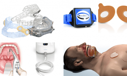
Satisfactory treatment of sleep disordered breathing can be achieved in almost all neuromuscular diseases with NPPV.6 Continuous positive airway pressure (CPAP) treatment is generally not indicated, as hypoventilation and central apnea are the predominant sleep pathology. It is recommended that the NPPV devices have a backup rate incorporated as central apneas can be quite prolonged.7 Individuals most often use nasal interfaces, although oronasal interfaces can be used if there is leak from the mouth or if the individual is more comfortable with the latter. Chinstraps also can be used if there is mouth leak. Occasionally, ventilation through an oral interface is used for patients with nasal problems preventing use of a nasal interface.8
NPPV treatment of sleep disordered breathing results in improvement of symptoms and has been shown to have a dramatic effect on survival in a number of different neuromuscular diseases.6 In addition, improvement and sometimes normalization of daytime PaCO2 can be achieved with nocturnal treatment. The exact mechanism of this daytime improvement is unknown, although prevention of recurrent elevations of PaCO2 at night may prevent retention of bicarbonate ions by the kidney or prevent resetting of respiratory drive centers in the central nervous system during hypercarbic and hypoxemic events.9
On occasion, patients will not tolerate NPPV at night. This may be because their neuromuscular disease has affected bulbar or facial muscles to the point where adequate ventilation without aspiration cannot be achieved. Sometimes patients do not wish or are unable to adapt to the facial interface despite desensitization and trials of multiple interfaces. In this case, tracheostomy will need to be considered because untreated sleep disordered breathing can lead to progressive respiratory failure and death.4 This is obviously a major decision that is not undertaken unless all other noninvasive options have been exhausted. In the past, negative pressure ventilation with devices such as the iron lung or cuirass ventilator was tried. Unfortunately, these devices were shown to increase obstructive events during sleep because of dyssynchrony with the activation of upper airway muscles by the patient and are rarely, if ever, used anymore.10
Seeing that sleep disordered breathing is common in patients with neuromuscular disorders, it is imperative to recognize the symptoms of this condition. The symptoms may be subtle and a high index of suspicion should be maintained in evaluating these types of patients. Routine testing of pulmonary function, respiratory muscle strength, and gas exchange in the clinic can be helpful in identifying individuals with a high likelihood of having sleep disordered breathing. Appropriate treatment with NPPV can result not only in improvement in sleep quality and symptoms but prevention of progressive respiratory failure as well.
Joshua Benditt, MD, is a professor of medicine at the University of Washington School of Medicine, Seattle. He is also director of respiratory care services, Northwest Assisted Breathing Center, University of Washington Medical Center. He can be reached at . Louis Boitano, MS, RRT, is codirector of the Northwest Assisted Breathing Center, University of Washington Medical Center. Boitano can be reached at .
REFERENCES
- Gibson G. Diaphragmatic paresis: pathophysiology, clinical features, and investigation. Thorax. 1989;44:960-970.
- Culebras A. Sleep and neuromuscular disorders. Neurol Clin. 1996;14:791-805.
- Barthlen GM, Lange DJ. Unexpectedly severe sleep and respiratory pathology in patients with amyotrophic lateral sclerosis. Eur J Neurol. 2000;7:299-302.
- Culebras A. Sleep and neuromuscular disorders. Neurol Clin. 2005;23:1209-1223, ix.
- Bach JR. Noninvasive respiratory muscle aids and intervention goals. In: Bach JR, ed. Noninvasive Mechanical Ventilation. Philadelphia: Hanley and Belfus; 2002:129-164.
- Mehta S, Hill NS. Noninvasive ventilation. Am J Respir Crit Care Med. 2001;163:540-577.
- Clinical indications for noninvasive positive pressure ventilation in chronic respiratory failure due to restrictive lung disease, COPD, and nocturnal hypoventilation—a consensus conference report. Chest. 1999;116:521-534.
- Bach JR, Alba AS, Saporito LR. Intermittent positive pressure ventilation via the mouth as an alternative to tracheostomy for 257 ventilator users. Chest. 1993;103:174-182.
- Misuri G, Lanini B, Gigliotti F, et al. Mechanism of CO(2) retention in patients with neuromuscular disease. Chest. 2000;117:447-453.
- Bach JR, Penek J. Obstructive sleep apnea complicating negative-pressure ventilatory support in patients with chronic paralytic/restrictive ventilatory dysfunction. Chest. 1991;99:1386-1393.
